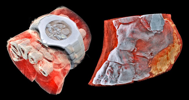
The new X-ray unit allows you to see the internal organs "in color"
Specialists of the New Zealand company Mars Bioimaging have created a medical device that allows to carry out the procedure of color 3D X-rays.
Classic black-and-white X-rays allow to diagnose many diseases, this method of research is especially indicative in determining fractures and other injuries of the skeletal system. The emergence of color images brought medical research to a new level.
The essence of the device is reduced to a special radiation of the body. The length of these waves allows you to penetrate the fabric. At the same time through the bone tissue waves can not penetrate. As a result, the resulting image is obtained two-dimensional.
Specialists from Mars Bioimaging used a similar technology, borrowing the principles of the Large Hadron Collider. The device's microcircuit works in approximately the same way as the sensors of digital cameras, the difference lies in the special method of fixing the particles of light when they hit the pixels.
The new modification of the chip makes it possible to detect fluctuations in wavelengths at the time of their passage through various substances of the body, be it bones, muscles, fluids, or other objects. Specialized software translates the incoming signals into a color and three-dimensional image. It looks like a three-dimensional photograph. Such X-rays can diagnose not only traditional fractures, cracks and displacements, but also other pathologies that are not visible to the ordinary X-ray eye.
/rating_off.png)